What is Ultrasound?
Ultrasound imaging, also called sonography imaging or ultrasound scanning, is a technique for getting pictures from inside the human body using high-repeat sound waves. The reflected sound wave echoes are recorded and shown as a consistent visual picture.
Be Prepared
Why is an Ultrasound Performed?
Don’t be Surprised!
How is an Ultrasound Performed?
The steps you will take to prepare for an ultrasound will depend on the area or organ that is being examined.
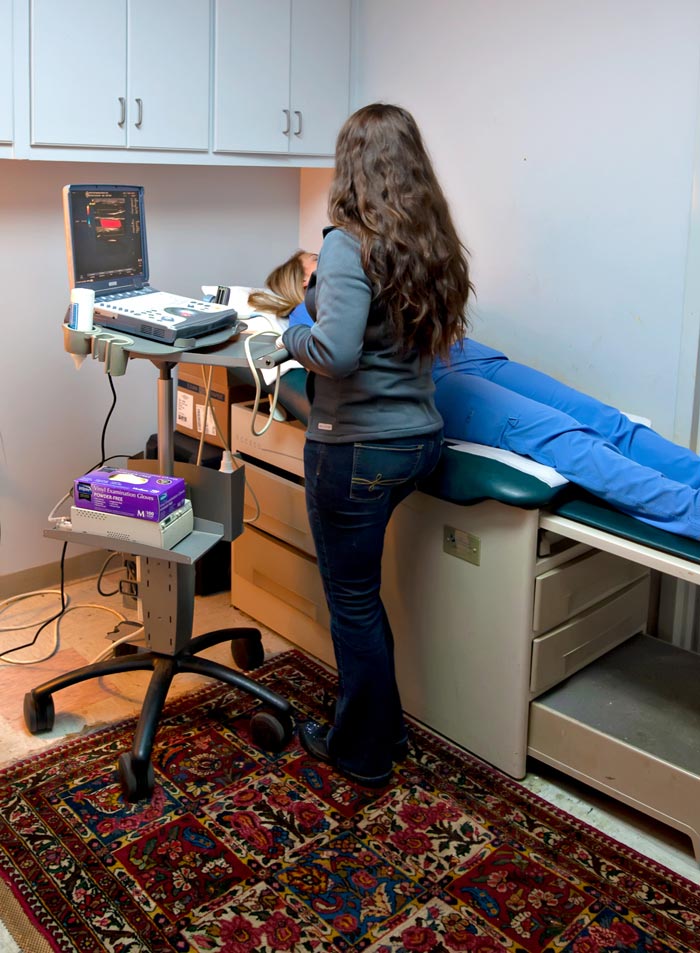
Before the test, you will change into a medical clinic gown. Then you will lie on your back on an examining table. An ultrasound expert will apply a special lubricating gel to your skin. This prevents friction so they can rub the ultrasound transducer on your skin. The gel also helps transmit sound waves. The reflected sound wave echoes are recorded and displayed as a real-time visual image.
After the procedure, the gel will be cleaned off of your skin. Ultrasound examinations usually take less than 30 minutes. You will be free to go about your normal activities after the procedure has finished.
We Will Take Care of The Rest!
What About the Results?
You will be free to leave the facility and resume normal activities as your health permits. A radiologist reads your sonogram, and the results are reported directly back to your doctor.


